

The human heart is a vital organ that pumps blood throughout the body. It is located in the chest and is roughly the size of a fist. The heart is made up of four chambers: the right atrium, right ventricle, left atrium, and left ventricle. Blood enters the heart through the right atrium and is pumped to the lungs for oxygenation by the right ventricle. Oxygen-rich blood then returns to the left atrium and is pumped out to the body by the left ventricle. The heart beats approximately 60-100 times per minute in a healthy adult.
Introduction to the Human Heart
The human heart is a muscular organ that is responsible for pumping blood throughout the body. It is located in the chest, between the lungs, and is roughly the size of a closed fist. The heart is a vital organ that is essential for the survival of an individual, as it supplies oxygen and nutrients to the body’s tissues and organs. It is made up of four chambers, including the right atrium, right ventricle, left atrium, and left ventricle. Blood enters the heart through the right atrium and is then pumped to the lungs for oxygenation by the right ventricle. Oxygen-rich blood returns to the left atrium and is then pumped out to the body by the left ventricle. The heart also has its own blood supply through the coronary arteries, which deliver oxygen and nutrients to the heart muscle itself. The heart is regulated by the autonomic nervous system and the sinoatrial node, often called the “natural pacemaker,” which initiates the heart’s rhythmic contractions.
Position of Heart in Human Body

The human heart is located in the chest, slightly to the left of the center of the chest. It is positioned behind the sternum (breastbone) and between the lungs. The base of the heart is at the top, where the major blood vessels enter and exit, and the apex is at the bottom, pointing toward the left hip. The heart is tilted slightly to the left, and about two-thirds of it is located to the left of the midline of the body, while the remaining one-third is located to the right of the midline. The exact position of the heart can vary slightly depending on the individual’s body size, shape, and posture.
Function of Heart
- Pumps blood throughout the body
- Delivers oxygen and nutrients to the body’s tissues and organs
- Removes waste products, such as carbon dioxide, from the body’s tissues and organs
- Regulates blood pressure
- Ensures that blood flows in the correct direction through the circulatory system
- Circulates white blood cells throughout the body, which helps fight infection and disease
- Plays a crucial role in maintaining homeostasis (balance) within the body
- Responds to the body’s demands, such as during exercise, by adjusting the heart rate and cardiac output to meet the body’s needs.
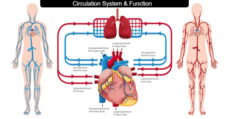
Types of Circulation
There are two types of circulation in the human body: pulmonary circulation and systemic circulation.
Pulmonary Circulation: This type of circulation involves the movement of blood between the heart and lungs. Deoxygenated blood is pumped from the heart’s right ventricle to the lungs, where it picks up oxygen and releases carbon dioxide. The oxygen-rich blood then returns to the heart’s left atrium, ready to be pumped out to the body.
Systemic Circulation: This type of circulation involves the movement of oxygenated blood from the heart to the body’s tissues and organs, and the return of deoxygenated blood to the heart. Oxygen-rich blood is pumped from the heart’s left ventricle to the rest of the body through the systemic arteries. The oxygen is delivered to the body’s tissues and organs, and the deoxygenated blood is then returned to the heart through the systemic veins, entering the right atrium. The right atrium then pumps the deoxygenated blood to the lungs to be oxygenated in the process of pulmonary circulation.
Structure of Human Heart
The human heart is a muscular organ that is roughly the size of a closed fist. It is made up of four chambers: the right atrium, right ventricle, left atrium, and left ventricle.
External Structure of Heart :-
The external structure of the human heart includes the following features:
- The heart is roughly cone-shaped, with a rounded apex and a broad base.
- The base of the heart is located at the top, where the major blood vessels enter and exit.
- The apex of the heart is located at the bottom and points toward the left hip.
- The heart is surrounded by a double-layered sac called the pericardium, which helps protect and lubricate the heart.
- The pericardium attaches to the diaphragm and the great vessels of the heart, helping to anchor the heart in place.
- The heart is located in the chest, between the lungs, and is slightly tilted to the left.
- The heart is roughly the size of a closed fist, with an average weight of about 250-350 grams in adults.
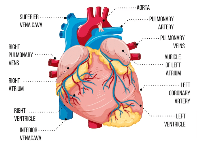
- The major blood vessels that enter and exit the heart are the superior and inferior vena cava, the pulmonary artery, and the aorta.
- The coronary arteries, which supply the heart muscle with oxygen and nutrients, also originate from the aorta.
- The heart is divided into four chambers, which are separated by valves and connected by blood vessels.
- The right side of the heart receives deoxygenated blood from the body and pumps it to the lungs for oxygenation, while the left side of the heart receives oxygenated blood from the lungs and pumps it to the rest of the body.
Pericardium
The pericardium is a double-layered sac that surrounds and protects the heart. The outer layer is called the fibrous pericardium, and it is tough and fibrous, while the inner layer is called the serous pericardium, which is a thin, smooth membrane that produces a lubricating fluid to reduce friction between the heart and surrounding structures. The pericardium also helps to anchor the heart in place within the chest and prevent it from over-expanding during periods of high blood flow. It is important for maintaining the structural integrity of the heart and facilitating its normal function.
Structure of the Heart Wall
The wall of the human heart is composed of three distinct layers, from outer to inner:
Epicardium: The outermost layer of the heart wall, also known as the visceral pericardium. It is a thin layer of connective tissue that covers the surface of the heart and helps reduce friction between the heart and surrounding structures.
Myocardium: The middle layer of the heart wall, composed of cardiac muscle tissue. The myocardium is responsible for the heart’s ability to contract and pump blood throughout the body. It is the thickest layer of the heart wall, and its thickness varies depending on the chamber it surrounds.
Endocardium: The innermost layer of the heart wall, composed of a thin layer of endothelial cells. The endocardium lines the interior surface of the heart and is continuous with the endothelial lining of blood vessels.
Internal Structure of Heart :-
The internal structure of the human heart is composed of four chambers, valves, and blood vessels. The chambers of the heart include:
Chambers of the Heart
The chambers are separated by valves and connected by blood vessels.
- Right atrium: Receives deoxygenated blood from the body through the superior and inferior vena cava.
- Right ventricle: Pumps deoxygenated blood to the lungs through the pulmonary artery.
- Left atrium: Receives oxygenated blood from the lungs through the pulmonary veins.
- Left ventricle: Pumps oxygenated blood to the body through the aorta.
Blood Vessels
Blood vessels are essential for the distribution of oxygen, nutrients, and other vital substances throughout the body, as well as the removal of waste products. The structure and function of blood vessels are highly regulated by the nervous and endocrine systems to ensure efficient blood flow and maintain normal blood pressure. Blood vessels are tubular structures that carry blood throughout the body. The three main types of blood vessels are:
Arteries: These blood vessels carry oxygenated blood away from the heart and distribute it to the body’s tissues and organs. Arteries have thick walls made of three layers: the tunica adventitia (outer layer), the tunica media (middle layer), and the tunica intima (inner layer). The middle layer is the thickest and is composed of smooth muscle, which helps regulate blood pressure.
Veins: These blood vessels carry deoxygenated blood back to the heart from the body’s tissues and organs. Veins have thinner walls than arteries and are less muscular. They also contain valves that help prevent the backflow of blood. Veins are divided into three layers: the tunica adventitia, the tunica media, and the tunica intima.
Capillaries: These tiny blood vessels connect arteries and veins and allow for the exchange of oxygen, carbon dioxide, nutrients, and waste products between the blood and the body’s tissues. Capillaries are composed of a single layer of endothelial cells, which are thin enough to allow for the diffusion of these molecules.
Also Check: Blood
Valves
Valves are structures within the circulatory system that help to regulate blood flow and prevent backflow. In the heart, there are four valves, two atrioventricular (AV) valves and two semilunar valves.
The two AV valves are the tricuspid valve and the mitral valve, which are located between the atria and ventricles. The tricuspid valve has three cusps, or leaflets, and is located on the right side of the heart. The mitral valve has two cusps and is located on the left side of the heart. These valves open to allow blood to flow from the atria into the ventricles, and then close to prevent the backflow of blood when the ventricles contract.
The two semilunar valves are the pulmonary valve and the aortic valve, which are located between the ventricles and the major arteries leaving the heart. The pulmonary valve is located between the right ventricle and the pulmonary artery, while the aortic valve is located between the left ventricle and the aorta. These valves have three cusps each and open to allow blood to flow out of the ventricles into the arteries, and then close to prevent the backflow of blood when the ventricles relax.
Valves are essential for maintaining the unidirectional flow of blood through the heart and circulatory system, and they are subject to wear and tear over time. Valvular disorders, such as stenosis or regurgitation, can result in impaired blood flow and can be managed with medications or, in some cases, surgical intervention.
Facts about Human Heart
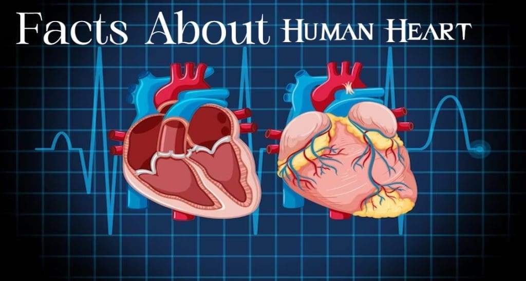
Here are some interesting facts about the human heart:
- The heart is about the size of a closed fist and weighs about 10-12 ounces in the average adult.
- The heart beats about 100,000 times per day, or around 3 billion times over the course of a lifetime.
- The heart pumps around 2,000 gallons of blood through the body each day.
- The heart is the first organ to develop in the human body, starting to form just 3 weeks after conception.
- The heart is located in the center of the chest, between the lungs and just behind the sternum (breastbone).
- The heart is composed of four chambers: the right atrium, right ventricle, left atrium, and left ventricle.
- The heart is nourished by its own blood vessels, known as the coronary arteries.
- The heart is surrounded by a fluid-filled sac called the pericardium, which helps to protect and lubricate the heart.
- The heart’s electrical system controls the heartbeat and is regulated by a group of specialized cells called the sinoatrial (SA) node.
- Heart disease is the leading cause of death globally, responsible for around 1 in 4 deaths.
Human Heart Drawing
Here knowledge glow provide drawing of human heart, human heart images, human heart drawing.


Frequently Asked Questions (FAQs)on Human Heart
What is the function of the heart?
The main function of the heart is to pump blood throughout the body to deliver oxygen and nutrients to the tissues and organs.
How many chambers does the heart have?
The heart has four chambers: the right atrium, right ventricle, left atrium, and left ventricle.
What is the pericardium?
The pericardium is a fluid-filled sac that surrounds and protects the heart.
What is a heart attack?
A heart attack occurs when blood flow to a part of the heart is blocked, usually by a blood clot, which can cause damage to the heart muscle.
What is high blood pressure?
High blood pressure, or hypertension, is a condition in which the force of blood against the walls of the blood vessels is too high, which can lead to cardiovascular problems.
What are some risk factors for heart disease?
Some common risk factors for heart disease include smoking, high blood pressure, high cholesterol, obesity, diabetes, and a family history of heart disease.
How can I keep my heart healthy?
To keep your heart healthy, you can maintain a healthy weight, exercise regularly, eat a balanced and nutritious diet, avoid smoking, and manage stress levels.
What is arrhythmia?
Arrhythmia is a condition in which the heart beats irregularly, either too fast or too slow.
What is heart failure?
Heart failure occurs when the heart is unable to pump enough blood to meet the body’s needs.
What is coronary artery disease?
Coronary artery disease is a condition in which the arteries that supply blood to the heart become narrow or blocked, which can lead to chest pain, heart attack, or other complications.


Recent Comments